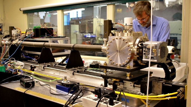
Mark LeGros, who spent three years building the world's first x-ray microscope dedicated to cell biology, greeted me at the Lawrence Berkeley National Laboratory (LBNL) with a hearty handshake, effusive smile and an accent reminiscent of his New Zealand roots.
I spent an hour in April talking with him about the challenges and rewards of creating a powerful, state-of-the art microscope which can capture a truer, more accurate picture of a whole cell and its nucleus, mitochondria and other tiny, essential cellular structures.
I asked him to take me back to that moment in 2009 when he and the diverse team of researchers, including biologists, physicists, chemists and computer scientists, who comprise the National Center of X-ray Tomography at LBNL, powered up the microscope and waited anxiously for the first x-ray images of a yeast cell, the biological specimen they chose to test the capabilities of their new imaging tool.

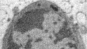
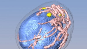
"I wasn’t sure exactly just how well this was gonna work out. But it turned out that it was beautiful...and everybody associated with the project was stunned with the degree of detail that it revealed about the internal structure of a cell and the chromatin, which is the genetic material of the cell," he said.
It's important to note that this powerful device does not render obsolete other imaging techniques, including light microscopy and electron microscopy. For one thing, the cell sample is frozen to hold the structures in place and protect them from the x-rays which have much greater penetrating power than visible light because x-rays have shorter wavelengths than light rays. So it can't capture dynamic activity of cells moving in real-time, unlike an optical or light microscope. Also, while the resolution possible with the x-ray microscope is five times greater than a light microscope, an electron microscope, which uses electrons to penetrate ultra-thin sections of cells, provides better resolution than the x-ray microscope.
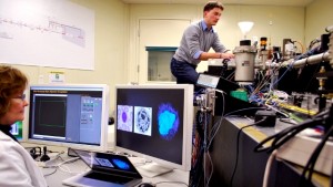
Nonetheless, unlike an electron microscope, the x-ray microscope allows scientists to image a whole cell in its native state, with the only preparation being the freezing of the cell sample. Also, many of the classic textbook images of cells that were taken with electron microscopes which required the cells to be dehydrated and stained with heavy metals to bind the electrons to the cell and generate a rather grainy image. Since a cell is 70% water, dehydration and staining can degrade delicate cellular structures and thus make it hard to accurately visualize them.
