We often look at skeletons (when we look at them at all) as reconstructions: bones dug out of the ground or dissected out of a body, then carefully fitted and wired back together. But this process can lose the skeleton’s natural orientation, leaving scientists to wonder how it would really look inside the animal. A vivid technique called clearing and staining offers just that perspective.
As a side effect, cleared and stained skeletons are strikingly beautiful. But not many people outside the lab would ever know it—until now.
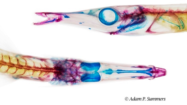
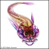
“Cleared” is an exhibit of stained fish skeletons currently on display at the Seattle Aquarium. The fish were prepared and photographed by Adam P. Summers, a biology professor at the University of Washington’s Friday Harbor Labs who also holds a research associate position at UC Berkeley. He uses the images for his research in biomechanics, a field that blends physics and biology.
To visualize a fish by clearing and staining, the body must first be fixed in formalin, then dehydrated in alcohol. The scientist-cum-artist then alternately stains the parts he wants to see (blue for cartilage, red for bone) and clears the parts that get in the way (bleach takes care of pigments, enzymes digest everything else). The result is a 3D view of the skeleton as it naturally fits together inside the body.
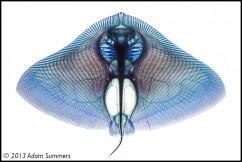
Such a view can offer scientific insight into otherwise intractable questions. Recently, Summers and his colleagues used a cleared and stained devil ray (a member of the group that includes manta rays) to discover how these curiously flat fish filter food out of the water.